|
|
|
|
Research Article |
The
Efficacy of Chest X-Ray for the Diagnosis and Follow-up in Young Adult
COVID-19 Patients with Mild Dyspnea No Comorbid Diseases Hafif Dispneli Komorbid Hastalığı Olmayan Genç Yetişkin
COVID-19 Hastalarında
Göğüs Röntgeninin Tanı ve Takipteki
Etkinliği *Ferhat CÜCE1 [ID], Deniz
DOĞAN2 [ID], Uğur
BOZLAR1 [ID], Mustafa
TAŞAR1 [ID], Abstract We investigated whether Chest X-Ray (CXR) could
replace computed tomography (CT) modality in the diagnosis and during the
treatment of young adult COVID-19 patients with mild dyspnea with no comorbid
diseases. This retrospective study involved an examination of the records of
a total of 956 patients hospitalized between March 1, 2020, and May 15, 2020.
The study included a total of sixty-four COVID-19 patients who underwent a
CXR at admission and CT imaging within 24 hours, aged 21-60 years with mild
dyspnea with no comorbid diseases. The diagnosis of infection was confirmed
by the polymerase chain reaction test in all cases. The first CXR and CT
images at the time of admission were evaluated in terms of lesions and
localization. The clinical-radiological course of the disease was also
statistically evaluated. CT was normal in 18/64 (28.1%) patients, all of whom
also had normal CXR. The rest of the patients 46/64 (71.9%) with an abnormal
CT, the CXR was normal in 18/46 (39.1%) and abnormal in 28/46 (60.9%). The
time between the onset of complaints and admission to the hospital in
patients with abnormal and normal CXR was 3.5±2.3 days and 2.1±1.1 days respectively and this difference was
statistically significant (p=0.004). The hospital stay durations of the
patients with abnormal and normal CXR was 9.6±3.5 and 9.5±3.4 days
(p=0.928), respectively, and was not statistically significant. In
conclusion, in the case of early admission to the hospital, there is not a
significant difference between using CXR or CT in the management of young
adult COVID 19 patients with mild dyspnea no comorbid disease. Therefore, the
use of CXR in these patient groups will reduce the burden of CT units in
pandemic conditions with limited resources. Keywords:
Chest X-ray, Ground-glass
opacity, Computed tomography, COVID-19. Özet Bu retrospektif çalışmada, komorbid
hastalığı bulunmaksızın hafif nefes darlığı olan genç erişkin COVID-19
hastalarının tanı ve tedavileri sırasında göğüs röntgeninin
bilgisayarlı
tomografi (BT) yönteminin
yerini alıp
alamayacağını araştırdık. Çalışmada 1 Mart 2020 ile 15 Mayıs 2020 tarihleri
arasında hastaneye yatırılan toplam 956 hastanın kayıtları
incelenmiştir. Çalışmaya,
kabul sırasında göğüs röntgeni çekilen ve 24 saat içerisinde BT görüntülemesi yapılan, 21-60 yaşları arasında hafif nefes
darlığı olan ve komorbid hastalığı olmayan toplam altmış dört
COVID-19 hastası dahil
edildi. Tüm
vakalarda enfeksiyon tanısı polimeraz zincir reaksiyonu testi ile
doğrulandı. Başvuru anındaki ilk göğüs röntgeni
ve BT görüntüleri
lezyonlar ve lokalizasyon açısından değerlendirildi. Hastalığın
klinik-radyolojik seyri de istatistiksel olarak değerlendirildi. Hastaların 18/64’ünde (%28.1) BT normaldi ve hepsinde
normal göğüs röntgeni vardı. Hastaların geri kalanı (46/64; %71.9) anormal BT'ye sahipti
ve göğüs röntgeni raporları bu hastaların 18/46'sında (%39.1) normal ve
28/46'sında
(%60.9) anormaldi. Anormal ve normal göğüs röntgeni olan hastalarda şikayetlerin başlaması ile hastaneye başvuru
arasındaki süre sırasıyla 3.5±2.3 gün ve 2.1±1.1 gün olup fark istatistiksel olarak anlamlıydı
(p=0.004). Anormal ve normal göğüs röntgeni olan hastaların hastanede yatış süreleri sırasıyla 9.6±3.5 ve 9.5±3.4 (p=0.928) gün idi ve istatistiksel olarak anlamlı değildi.
Sonuç olarak, hastaneye erken başvuru
durumunda hafif dispnesi olan ve komorbid hastalığı olmayan genç erişkin
COVID-19 hastalarının yönetiminde
göğüs röntgeni
veya BT kullanımı arasında anlamlı bir
fark yoktur. Bu nedenle, bu hasta gruplarında göğüs röntgeni
kullanımı,
sınırlı kaynaklarla pandemi koşullarında BT ünitelerinin yükünü azaltacaktır. Anahtar kelimeler: Göğüs röntgeni, Buzlu cam
opasitesi, Bilgisayarlı tomografi, COVID-19.
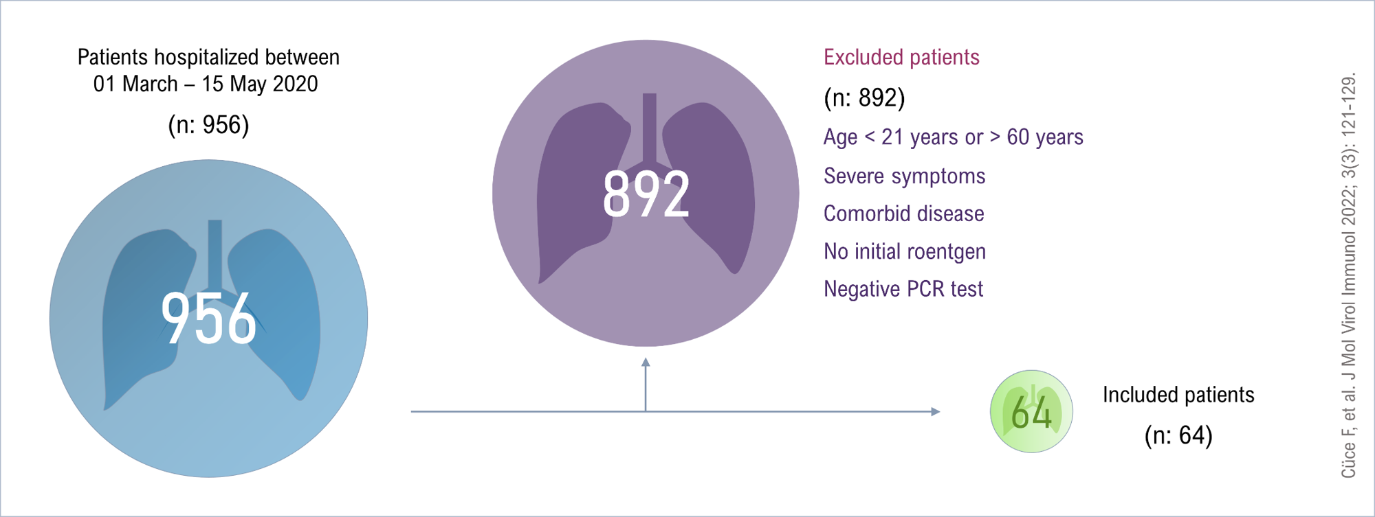
Figure 1. Flow of
diagram of the retrospective study (PCR: Polymerase Chain Reaction). Figure 1 png Şekil 1. Retrospektif çalışmanın akış diyagramı
(PCR: Polimeraz Zincir Reaksiyonu). Şekil 1 png
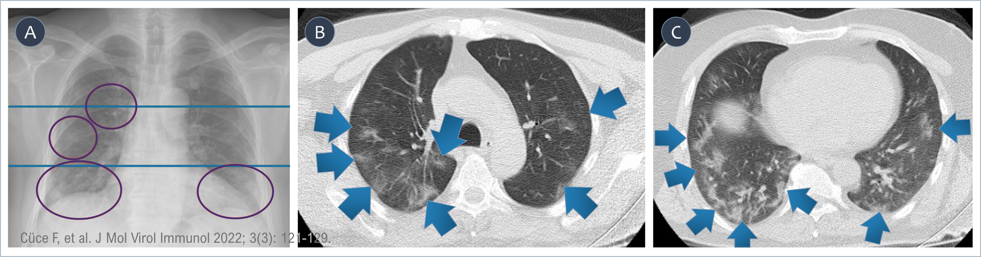
Figure 2. A 43-year-old male patient with COVID 19 and without any comorbid
disease presenting with mild dyspnea. (A) In CXR, faint-border
opacities are observed in the right upper and middle zone and bilateral lower
zones. Thoracic CT in the axial plane shows central and peripherally
localized ground glass density lesions in bilateral upper (B) and lower zones (C). Figure 2 png Şekil 2. Hafif nefes darlığı
şikayetiyle başvuran, herhangi bir komorbid hastalığı olmayan, 43 yaşında,
COVID 19 tanılı erkek hasta. (A) In CXR, faint-border opacities are observed in the right upper and
middle zone and bilateral lower zones. CXR'de sağ üst ve orta zonda ve iki
taraflı alt zonda silik sınır opasiteleri izleniyor. Aksiyel düzlemdeki
toraks BT'de, her iki tarafta üst (B) ve alt (C) zonlarda merkezi ve periferik lokalize buzlu cam yoğunluğu lezyonları
görülüyor. Şekil 2 png
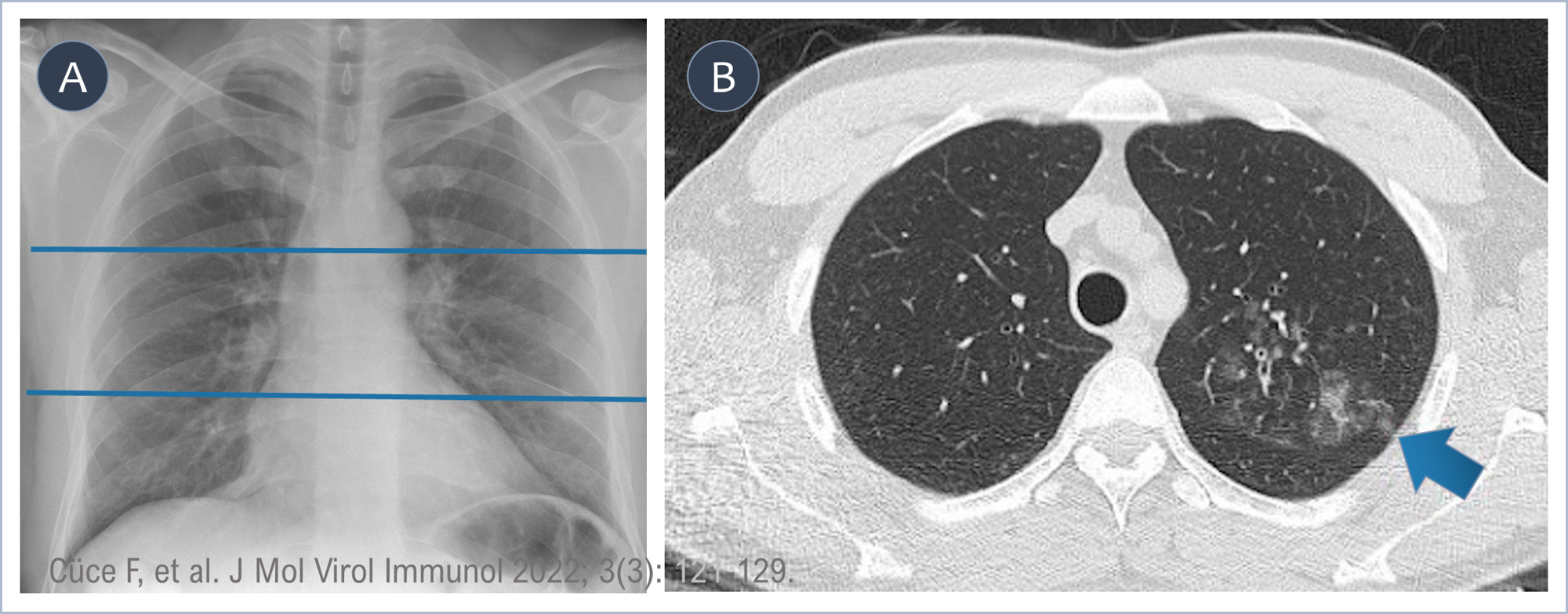
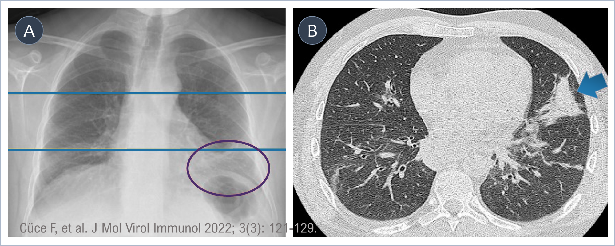
Figure 3. A 23-year-old
male patient with COVID 19 complaining of mild dyspnea. (A) CXR is
false-negatively normal. (B) Ground-glass densities compatible with active infiltration are
observed in the left upper zone in the axial plane of thoracic CT. Figure 3 png Şekil 3. Hafif nefes darlığından şikayetçi olan 23
yaşında COVID 19'lu erkek hasta. (A) CXR yanlış negatif olarak normaldir. (B) Toraks BT'nin aksiyal düzleminde
sol üst zonda aktif infiltrasyonla uyumlu buzlu cam yoğunlukları gözleniyor. Şekil 3 png
Figure 4. A 34-year-old
male patient with COVID 19 complaining of mild dyspnea. Şekil 4. Hafif nefes darlığından şikayetçi olan 34
yaşında COVID 19 tanılı erkek hasta.
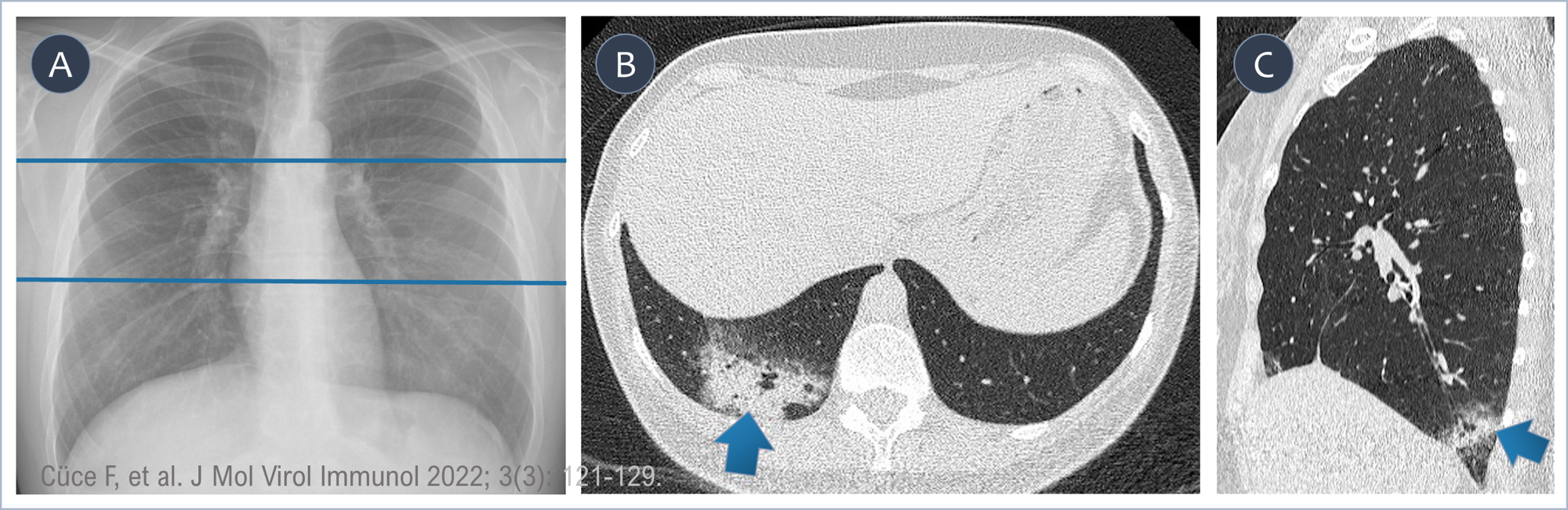
Figure 5. A 38-year-old
female patient with COVID 19 complaining of mild dyspnea. (A) There is an
increased opacity in CXR in the lower-left zone. (B) The homogeneous hyperdense
consolidated atelectatic parenchyma in the lower left zone in the axial plane
of the thorax CT is consistent with the opacity in the CXR. While the heart
and diaphragm can obscure GGO-type lesions, consolidation, which is one of
the later signs of the disease, can be detected with CXR due to its more
opaque appearance. Figure 5 png Şekil 5. Hafif nefes darlığından şikayetçi olan 38
yaşında COVID 19'lu kadın hasta. (A) CXR'de sol alt bölgede artan bir opaklık var. (B) Toraks BT'nin
aksiyel düzleminde sol alt zondaki homojen hiperdens konsolide atelektazik
parankim, CXR'deki opasite ile uyumludur. Kalp ve diyafram GGO tipi
lezyonları gizleyebilirken, hastalığın geç belirtilerinden biri olan
konsolidasyon, daha opak görünümü nedeniyle CXR ile tespit edilebilmektedir. Şekil 5 png |
|
DOI: 10.46683/jmvi.2022.56 |
|
|
Article in English |
|
|
|
|
|
|
|
|
1Department of Radiology, Gulhane Training
and Research Hospital, University of Health Sciences, Ankara, Türkiye. 2Department of Pulmonology, Gulhane
Training and Research Hospital, University of Health Sciences, Ankara, Türkiye. 3Department of Infectious Disease,
Gulhane Training and Research Hospital, University of Health Sciences,
Ankara, Türkiye. 4Department of Family Medicine, Gulhane
Training and Research Hospital, University of Health Sciences, Ankara, Türkiye. |
|
|
|
|
|
*Corresponding author Ferhat Cüce; Assoc.Prof., Department
of Radiology, Gulhane Training and Research Hospital, University of Health
Sciences, Ankara, Türkiye. E-mail: ferhatcuce@hotmail.com |
|
|
|
|
|
Received: 29.05.2022 Accepted: 08.07.2022 Published: 14.07.2022 |
|
|
Cite as: Cüce F,
Doğan D, Bozlar U, Taşar M, Gjoni E, Ayaz T, Savaşçı Ü, Sarı O. The Efficacy
of Chest X-Ray for the Diagnosis and Follow-up in Young Adult COVID-19
Patients with Mild Dyspnea No Comorbid Diseases. J Mol Virol Immunol 2022;
3(3): 121-129. |
|
|
|
|
|
View in academic indexes and databases |
|
|
|
|
|
|
|
|
|
|
|
|
|
|
|
|
|
|
|
|
|
|
|
|
|
|
|
|
|
|
|
|
|
|
|
|
|
|
|
|
|
|
|
|
|
|
|
|
|
|
|
|
|
|
|
|
|
|
|
|
|
|
|
|
|
|
|
|
|
|
|
|
|
|
|
|
|
|
|
|
|
|
|
|
|
|
|
|
|
|
|
|
|
|
|
|
|
|
Cited by 0 article*, 0 book chapter. |
|
|
|
|
|
©Copyright JMVI.
Licensed by Creative Commons Attribution-NonCommercial 4.0 International (CC
BY-NC 4.0). |
|