|
|
|
|
Research Article |
Catching
the First Wave in the Pandemic: A Retrospective Evaluation of Chest CT Images
for COVID-19 Pandemide İlk Dalgayı Yakalamak: COVID-19 için Göğüs BT
Görüntülerinin Retrospektif Değerlendirilmesi *Mehmet
TAHTABAŞI1 [ID], Ergin KARAMAN1 [ID], Yasin AKIN1 [ID], Osman
KONUKOĞLU1 [ID], Nihat KILIÇASLAN1 [ID], Mehmet GEZER1 [ID], Sadettin ER2
[ID], Selda TEZ3 [ID], Mesut TEZ2 [ID], Kemal TEKİN4 [ID] Abstract Pandemics generally begin in small areas and subsequently spread
widely. In order to detect an outbreak in its early stage, observation of the
data for small regions is important. The aim of this study to investigate the
presence of COVID-19-like pneumonia findings in computed tomography (CT)
taken before the COVID-19 pandemic in Turkey. The chest CTs performed in our
hospital between December 1, 2019 and March 10, 2020
(study group) and those performed between December 1, 2018 and March 10, 2019
(control group) were retrospectively analyzed. A total of 1,432 chest CT
images in the study group and 1,443 chest CT images in the control group were
evaluated for COVID-19. The lesion characteristics on CT, length of hospital
stay (LOS), and mortality rate were investigated. Typical lung involvement
defined for COVID-19 was present in 1.39% (n=20/1,432) of CTs in the study
group and 0.49% (n=7/1,443) in the control group (p=0.011). Seventy-five
percent (n=15/20) of the study group were male, and the mean age of the
patients was 51.8 (±17.1) years. All the patients in the study group had at
least one of the symptoms of COVID-19, such as fever, cough, and respiratory
distress. Ninety percent (n=18/20) of the patients in the study group had
ground-glass opacities that showed a predominantly peripheral distribution.
Five of these had accompanying consolidation and one had a reverse halo sign.
According to clinical records, in-hospital mortality developed in seven of 20
patients (35%), the LOS was 5.5±6.2 days, and the median time from the
symptom onset to admission was 4 (range: 1-12) days. Our study reveals that
the onset of COVID-19 or a similar disease is more likely to occur earlier
than first reported in the country. Keywords:
COVID-19, Early detection,
Computed tomography, Epidemiology, Pandemic. Özet Pandemiler genellikle küçük alanlarda
başlar ve daha sonra geniş bir alana yayılırlar. Bir salgını erken aşamada
tespit etmek için küçük bölgelerdeki verilerin gözlemlenmesi önemlidir. Bu
çalışmanın amacı Türkiye'de COVID-19 pandemisinden önce çekilen bilgisayarlı
tomografi (BT) görüntülerinde COVID-19 benzeri pnömoni bulgularının varlığını
araştırmaktır. Hastanemizde 1 Aralık 2019-10 Mart 2020 (çalışma grubu) ve 1
Aralık 2018-10 Mart 2019 (kontrol grubu) tarihleri arasında yapılan göğüs BT'leri geriye dönük olarak
incelendi. Çalışma grubundaki toplam 1.432 göğüs BT görüntüsü ve
kontrol grubundaki 1.443 göğüs BT görüntüsü
COVID-19 için değerlendirildi. BT'de lezyon özellikleri, hastanede yatış süresi
(HYS) ve mortalite oranı araştırıldı. COVID-19 için tanımlanan tipik akciğer
tutulumu, çalışma grubundaki BT'lerin %1.39'unda (n=20/1,432) ve kontrol
grubunun %0.49'unda (n=7/1,443) mevcuttu (p=0.011). Çalışma grubunun yüzde
yetmiş beşi (n=15/20) erkek olup, hastaların yaş ortalaması 51.8’dir (±17,1).
Çalışma grubundaki tüm hastalarda ateş, öksürük ve solunum sıkıntısı gibi
COVID-19 semptomlarından en az biri vardı. Çalışma grubundaki hastaların
yüzde doksanı (n=18/20) ağırlıklı olarak periferik dağılım gösteren buzlu cam
opasitelerine sahipti. Bunlardan beşinde bu bulgulara konsolidasyon eşlik
ederken ve birinde ters hale işareti vardı. Klinik kayıtlara göre 20 hastanın
yedisinde (%35) hastane içi mortalite gelişti, HYS 5.5±6.2 gün ve semptom
başlangıcından başvuruya kadar geçen medyan süre 4 (aralık: 1-12) gündü.
Çalışmamız, COVID-19 veya benzeri bir hastalığın başlangıcının ülkemizde ilk
bildirilenden daha erken ortaya çıkma olasılığının yüksek olduğunu ortaya
koymaktadır. Anahtar kelimeler: COVID-19, Erken teşhis,
Bilgisayarlı tomografi, Epidemiyoloji, Pandemi.
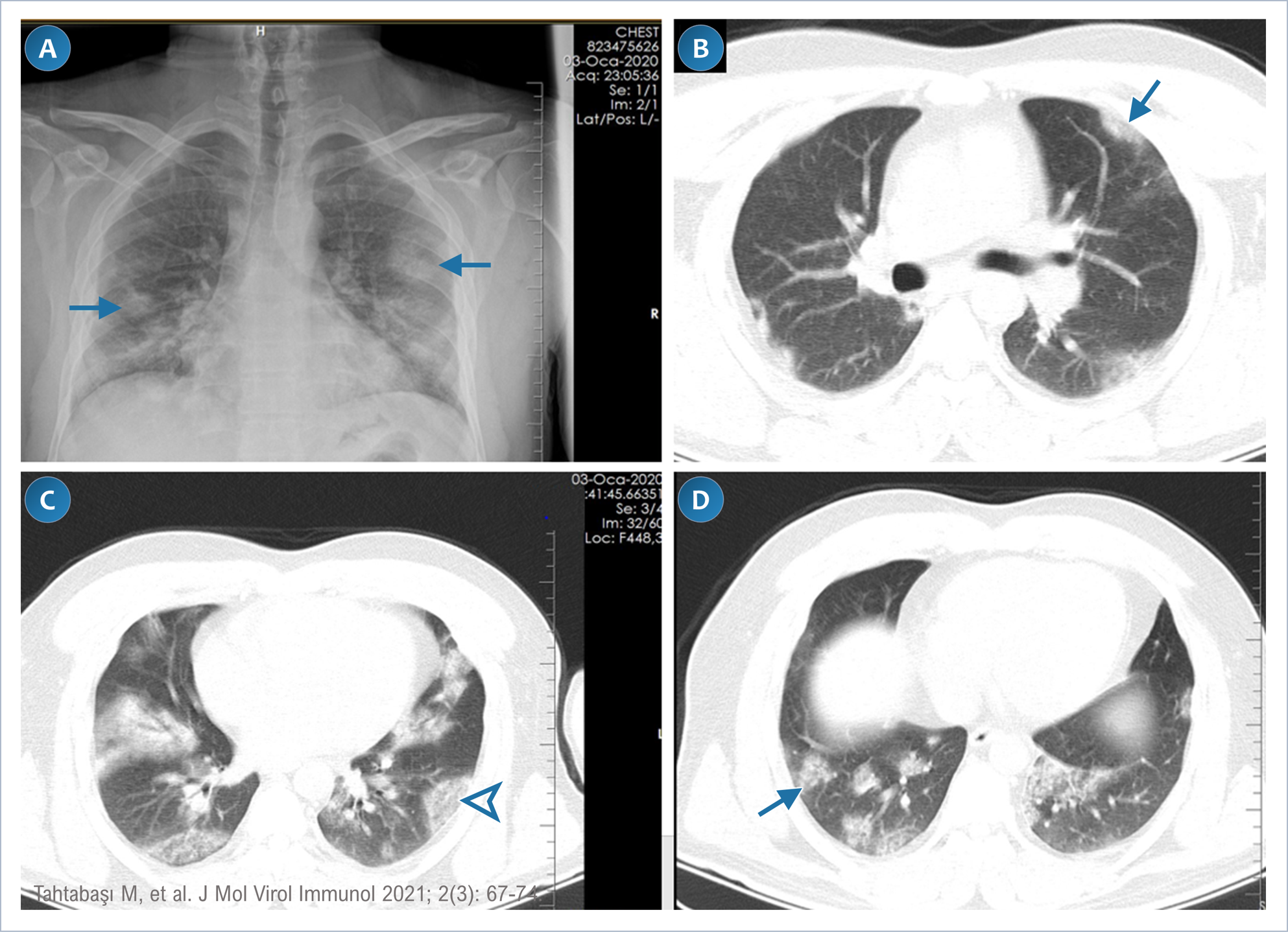
Figure 1. Chest X-ray and CT
images of a 33-year-old male patient taken in January 2020. X-ray image (A)
shows peripherally distributed opacities. CT images in the lung window (B,C,D)
shows peripherally distributed ground glass opacities, and the reverse halo
sign (arrowhead) with consolidated periphery and ground glass opacities in
the center. Figure 1 png Şekil 1. 33 yaşındaki erkek hastanın Ocak 2020'de çekilen göğüs
röntgeni ve BT görüntüleri. Röntgen görüntüsünde (A) periferik olarak
dağılmış opasiteler görülmekte. Akciğer penceresindeki (B,C,D) BT
görüntüleri, periferik olarak dağılmış buzlu cam opasitelerini ve konsolide
çevre ve merkezde buzlu cam opasiteleri ile ters hale işareti (ok başı)
göstermekte. Şekil 1 png
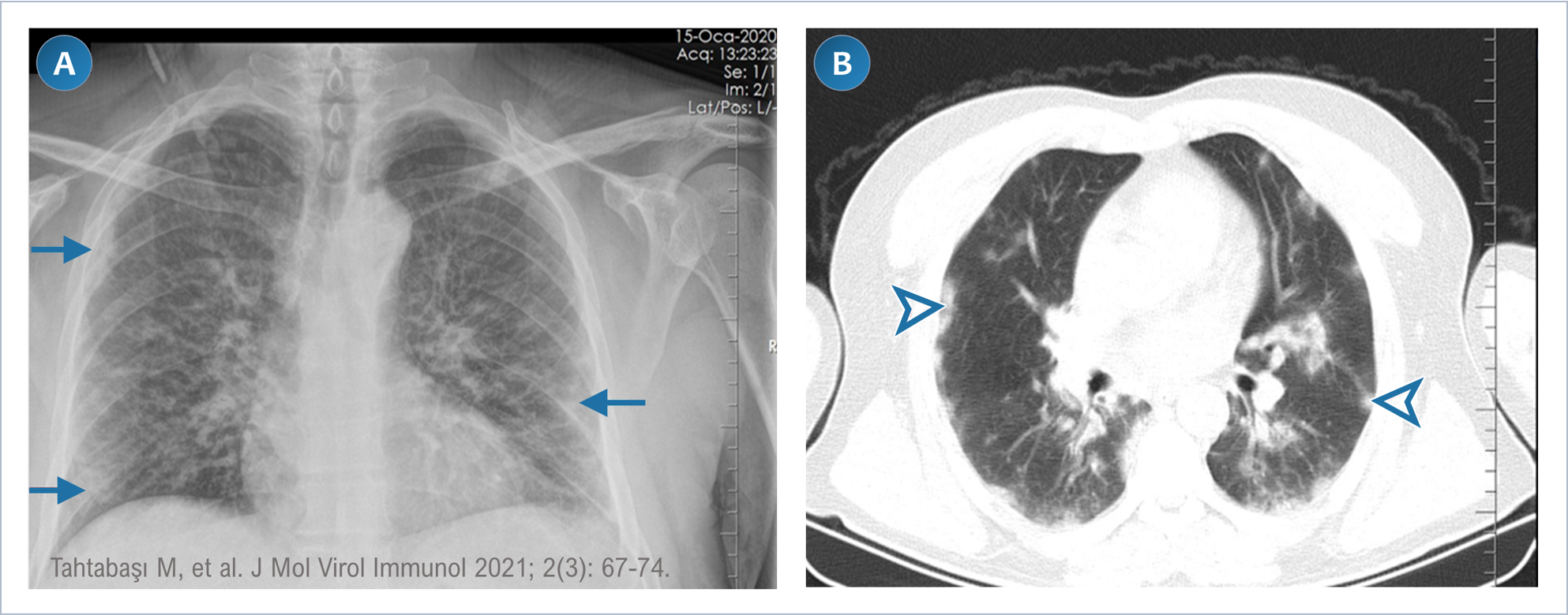
Figure 2. Chest X-ray and CT
images of the lung window of a 50-year-old male patient taken in January
2020. X-ray (A) and CT (B) images show peripherally distributed
ground glass opacities. Figure 2 png Şekil 2. 50 yaşındaki erkek hastanın Ocak 2020'de çekilen
akciğer penceresinin göğüs röntgeni ve BT görüntüleri. Röntgen (A) ve
BT (B) görüntüleri periferik olarak dağılmış buzlu cam opasitelerini
göstermekte. Şekil 2 png
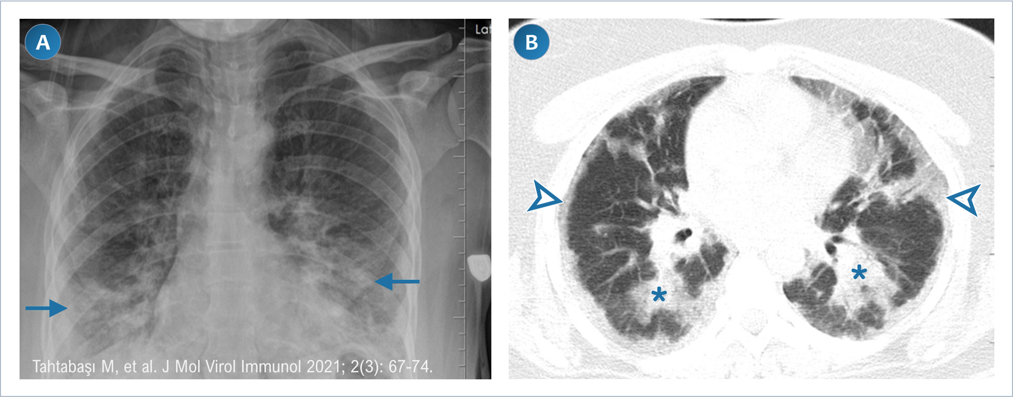
Figure 3. Chest X-ray and CT images in the lung window of a
39-year-old female patient taken in January 2020. (A) X-ray images show symmetrical opacities (arrows) in
the lower zones of the lung. (B) CT images show ground glass opacities (arrowheads)
located in the peripheral and subpleural parts of the lung and patchy
consolidations (asterisk) in the lower lobes. Figure 3 png Şekil
3.
39 yaşındaki kadın hastanın Ocak 2020'de çekilen akciğer penceresindeki göğüs
röntgeni ve BT görüntüleri. (A) Röntgen görüntülerinde akciğerin alt
zonlarında simetrik opasiteler (oklar) görülmekte. (B) BT
görüntülerinde akciğerin periferik ve subplevral
kısımlarında yer alan buzlu cam opasiteleri (ok başları) ve alt loblarda
yamalı konsolidasyonlar (yıldız) görülmekte. Şekil 3 png |
|
DOI: 10.46683/jmvi.2021.33 |
|
|
Article in English |
|
|
|
|
|
|
|
|
1Department of Radiology, Mehmet Akif
Inan Education and Research Hospital, University of Health Sciences, Şanlıurfa, Türkiye. 2Department of General Surgery, Ankara
City Hospital, Ankara, Türkiye. 3Department of Radiology, 19 Mayıs
Hospital, Ankara, Türkiye. 4Department of Medical Microbiology, Gulhane Training and Research Hospital, University of
Health Sciences, Ankara, Türkiye. |
|
|
|
|
|
*Corresponding author Mehmet Tahtabaşı;
Asst.Prof., Department of Radiology, Mehmet Akif
Inan Education and Research Hospital, University of Health Sciences, Şanlıurfa, Türkiye. E-mail: mehmet.tahtabasi@sbu.edu.tr |
|
|
|
|
|
Received: 11.06.2021 Accepted: 20.06.2021 Published: 21.06.2021 |
|
|
Cite as: Tahtabaşı M, Karaman E, Akın Y, Konukoğlu O, Kılıçaslan N, Gezer M, Er S, Tez S, Tez M,
Tekin K. Catching the First Wave in the Pandemic: A Retrospective Evaluation
of Chest CT Images for COVID-19. J Mol Virol
Immunol 2021; 2(3): 67-74. |
|
|
|
|
|
View in academic indexes and databases |
|
|
|
|
|
|
|
|
|
|
|
|
|
|
|
|
|
|
|
|
|
|
|
|
|
|
|
|
|
|
|
|
|
|
|
|
|
|
|
|
|
|
|
|
|
|
|
|
|
|
|
|
|
|
|
|
|
|
|
|
|
|
|
|
|
|
|
|
Cited by 1 article*, 0 book chapter. |
|
|
[1]* Bolat A, Cüce F, Şenoğlu MÇ, Şahiner A, Ünay B. Efficacy of Clinical and Chest Radiography
Features in Predicting Patient Prognosis in Children with COVID-19. J Mol Virol Immunol 2021; 2(4): 159-167. doi:
10.46683/jmvi.2021.43 |
|
|
©Copyright JMVI.
Licensed by Creative Commons Attribution-NonCommercial
4.0 International (CC BY-NC 4.0). |
|